


As a person ages, the risk of developing spine and joint diseases increases. This is due to degenerative and destructive changes in the body. One of the common pathologies is osteoarthritis of the ankle joint.
Arthrosis of the ankle joint: what is it?
Ankle osteoarthritis is a chronic disease and cannot be completely cured. According to statistics, 10% of people suffer from this dystrophic disorder. People over 40 are especially susceptible to it. The disease can cause disability. Therefore, it is necessary to treat it quickly and competently.
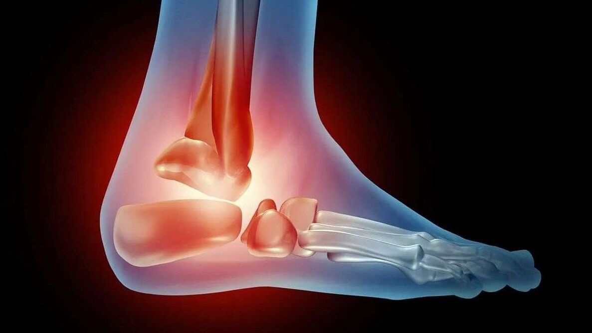
The ankle is formed by the fibula, talus and tibia, two malleoli and articular ligaments. With osteoarthritis, inflammation and destruction of articular cartilage occurs. The bone tissue is damaged and deformed as the pathology progresses.
ICD code 10
ICD stands for International Classification of Diseases. In this document, each disease is assigned a specific code. This code consists of letters and numbers and is indicated on the sick leave certificate when making a diagnosis. Thanks to it, a doctor from any country will be able to understand what the patient is suffering from and where the pathological focus is located.
The diagnosis of osteoarthritis is presented in a block of 5 titles and several subtitles. Ankle osteoarthritis is included in category M19. This section is divided into 5 subsections. The sign after the period indicates the etiology. So, 0 – these are genetically determined degenerative changes, 1 – post-traumatic changes, 2 – dystrophic changes against the background of endocrine, vascular or inflammatory pathology, 8 – these are other specified causes, 9 – a disease of unknown cause. For example, code M19. 1 is osteoarthritis of the ankle resulting from injury.
Causes
Pathology develops for several reasons. The factors that cause the appearance of the disease in adults are:
- Greater load on the joint. Doctors often see degenerative changes in cartilage and bone tissue in obese patients and in professional athletes (footballers, bodybuilders, runners, and dancers).
- Diabetes.
- Ankle injury.
- Wearing uncomfortable shoes, walking in heels.
In children, pathology develops for the following reasons:
- Thyrotoxicosis.
- tissue dysplasia.
- Injury.
- Genetic predisposition.
- Fracture.
- Joint inflammation.
- Dislocation.
Symptoms
The following manifestations are typical of ankle osteoarthritis:
- Pain. Appears after staying in a position. When a person tries to get up and lean on their leg, they experience stabbing pain (pain) and stiffness of movement. After a few steps the discomfort disappears. Pain appears during and after physical activity.
- Clicking, cracking in the ankle joint when walking.
- Limitation of movements.
- Swelling below the ankles.
- Hypotrophy, weakness of the ligamentous apparatus.
- Joint deformation (typical of advanced disease).

Degrees
There are various degrees of osteoarthritis. Many years pass from the appearance of the first signs of degenerative changes in the joint to the loss of mobility. If you start therapy on time, there is a chance to stop the progression of the disease. The success of treatment depends on the stage at which the pathology was detected.
Degrees of osteoarthritis of the ankle joint:
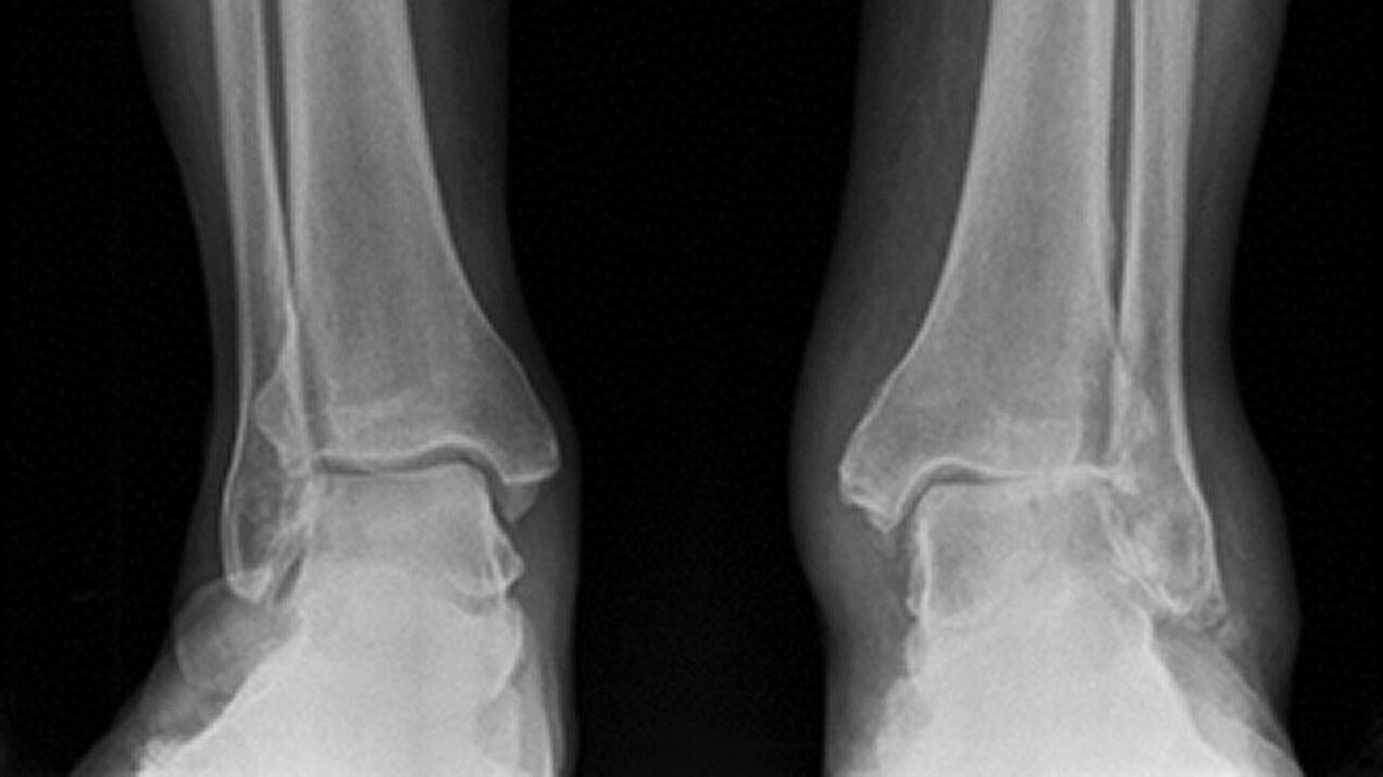
- First. The degenerative process is just beginning to develop and does not cause much discomfort to the person. The only symptoms are temporary morning stiffness in the legs, fatigue, and mild pain. When you bend and straighten your foot, a crunching sound is produced. No pathological changes are detected on the radiograph. The prognosis of pharmacological treatment is favorable.
- Second. The symptoms of the disease intensify. Morning stiffness doesn't go away for about an hour. The pain appears at the beginning of walking. Having covered only 1 km of distance, a person feels very tired in his legs. When the ankle moves, a cracking sound is produced. X-rays show osteophytes, the convergence of the ends of bones. Surgical treatment is indicated.
- Third. Pain syndrome occurs not only during movement, but also at rest. A person cannot work or rest normally without anesthetics. The patient cannot move independently. The X-ray image shows cracks, flattening of the articular surfaces, osteophytes and subluxations. The treatment is surgical and medicinal.
- Four. The manifestations of the disease are mild. The pain disappears. But the rigidity of movement does not allow a person to walk. The cartilage in the fourth stage is completely destroyed. The x-ray shows healing of the joint space.
Diagnosis
During diagnosis, the doctor determines the degree of the disease and identifies the exacerbation. For this, laboratory and hardware techniques are used:
- Blood test (detailed).
- Rheumatoid tests.
- Ultrasound.
- CONNECTICUT.
- PCR test.
- Bone scan.
- magnetic resonance.
Treatment
Therapy should be comprehensive and include taking medications, using physiotherapy methods, and performing therapeutic physical exercises.
The following medications are prescribed to the patient:
- Non-steroidal anti-inflammatory drugs.
- Chondroprotectors.
- Analgesics.
- Corticosteroid hormones.

Joint mobility is restored through manual therapy and procedures using a special device. Physiotherapy accelerates regeneration and stimulates blood circulation in the affected joint. Electrical stimulation, laser therapy, and ultrasound are effective. In case of pronounced dystrophic changes, endoprostheses are performed.
Prevention
You can prevent ankle osteoarthritis by following the following rules:
- Maintain weight within normal limits.
- Strengthen the spine with special exercises.
- Avoid injuries.
- Correct congenital anomalies of the joint structure.
- Quit smoking and drinking alcoholic beverages.
- Treat endocrine and vascular disorders in a timely manner.
- Periodically carry out preventive examinations if you have a genetic predisposition to the disease.